One of the greatest turning points in rhinoplasty over the last fifty years came when the focus switched from elimination to preservation and sometimes even augmentation. Still today, however, through carelessness or ignorance, many surgeons unfortunately continue to remove excessive amounts of the septum, dorsum, and alar and lateral cartilages and thus encounter the associated aesthetic and functional problems, including collapse of the nasal bridge, perforation of the septum, pinching of the nasal wings, saddle nose, and internal valve stenosis. Revision rhinoplasty is one of the most technically difficult operations in nasal surgery due to the existence of problems arising from previous surgery that make it more difficult to treat and handle the patient. On one hand, the bone and cartilage structures are weakened and distorted, and the soft, covering tissues are scarred and adherent. On the other hand, the patient often presents psychological problems, scepticism and diminished confidence in the surgeon. Revision rhinoplasty requires a combination of experience, architecture and art, as well as careful preoperative planning in order to restore balance between the functional and aesthetic aspects. All this entails a deep theoretical knowledge of the subject as codified in the literature and outstanding surgical dexterity. The use of the open approach has been proved as very useful in most cases, as it offers the enormous advantage of complete anatomical exposure. The far better view of the situation obtained with respect to the closed approach makes it possible to formulate a precise diagnosis. Another key advantage regards suturing and ensuring the stability of grafts. Revision rhinoplasty is often a work of actual architecture, aimed at rebuilding a structure that was damaged during previous surgery. Just as bricks must be laid with mortar to hold them in place, grafts must be firmly secured if the structure is to avoid collapse.

DROOPING TIP AND OTHER PROBLEMS
This patient, who had undergone two previous operations of rhinoplasty to remove a hump and correct crookedness of the nasal pyramid, complained of collapse of the lower section of the nose and persistence of the deviation as well as impaired nasal respiration.

Clinical analysis revealed deformity of profile caused by collapse of the lower third, deviation of the lower two-thirds of the nasal pyramid, marked deficiency of the septum, asymmetry of the tip, a drooping columella, and an acute nasolabial angle. The correction carried out through the open approach involved grafts of cartilage from the auricular concha. The placement of a spreader graft on either side of the septum corrected the deviation, a columellar strut graft provided new support for the lower third of the nasal pyramid, and another graft reconstructed the alar cartilage of the tip, which had been removed during previous surgery.

I published this case in 2010 in the article ‘Perichondrium Graft: Harvesting and Indications in Nasal Surgery,’ Journal of Craniofacial Surgery. 2010, 21, pp. 40–44.

POLLY BEAK DEFORMITY
This patient had previous undergone rhinoplasty to remove a hump and reshape the nasal tip, but the lower portion of the nose and septum remained crooked and his respiration was considerably impaired. His profile was also marred by an unsightly swelling in the area above the nasal tip.

Analysis of the patient revealed deviation of the nasal bridge, polly beak deformity of the profile, asymmetry of the nasal tip, and pinched nasal wings with excessive columellar show. These defects were corrected by removing an excess of the septum in the supratip region and through the placement of grafts of septal cartilage in precise nasal areas: a spreader graft on the right side of the septum to correct the deviation and two alar batten grafts to correct the pinching of the nasal wings.

A BOTTICELLI FACE
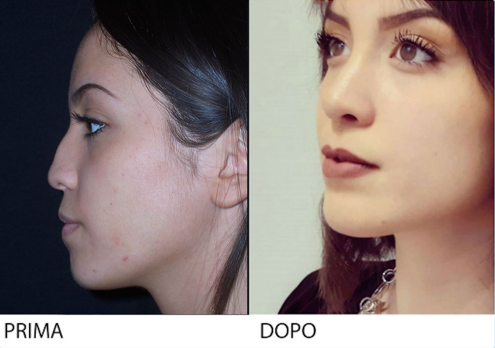
This patient, who had previous undergone septorhinoplasty, complained about an unsightly hump in the supratip area, a wide and drooping tip, and impaired nasal respiration, especially on the right nostril.

Analysis of the patient revealed the presence of polly beak deformity in the supratip region and a bulbous and poorly defined tip. Anterior rhinoscopy also revealed deviation of the nasal septum to the right. Rhinoplasty was performed with the open approach and, as usual, left no visible columellar scar. The lower third of the dorsum was lowered to bring it into line with the upper two-thirds and the tip was reshaped and rotated slightly upward. The patient’s delicate features suggested a more significant lowering of the bridge and greater rotation of the tip, so as to attain the end result of a Botticelli-like appearance, enhancing her beautiful and highly expressive eyes.

THE PSYCHOLOGICAL ASPECTS OF REVISION RHINOPLASTY
This patient complained of evident nasal blemishes as a result of two previous operations as well as impaired respiratory. During the interview he expressed his frustration with this unacceptable aesthetic situation, which also affected his relations with others.

Analysis of the patient revealed nasal deformity with flaws deriving from the removal and weakening of the cartilage structures of the bridge and the tip. The lateral walls of the nose were pinched, the tip excessively projected and upturned, and the supratip area unduly concave. Structural weakening was also responsible for malfunctioning of the nasal valve and impaired respiration above all during forced inspiration in moments of particular need. Revision rhinoplasty, performed with the open approach, used reconstructive grafts of cartilage from the auricular concha to give the patient’s nose a natural, uniform and overall compact appearance.

Patient Testimonial

“The morphology of my nose prior to the operation of revision rhinoplasty created a condition of existential and relational anxiety, especially in the working environment, which had gone on for years with repercussions in my private and professional life. My long and tormented clinical history regarding the septorhinoplasty began about eight years ago with a catastrophic operation at a hospital in Naples. The removal of cartilage caused the collapse of the nasal wall with both functional and aesthetic damage. One year later, I underwent electrosurgery to reduce the scarring and adherence hoping in a functional improvement at least, but without obtaining any benefit whatsoever. These failures only increased my frustration (the physicians who examined me were horrified but offered no solutions) and I lived in a state of impotence and hopelessness. After several years, it had become impossible for my wife to bear my constant snoring with intervals of authentic sleep apnea. She finally overcame my resistance and persuaded me to consult an ear, nose and throat specialist in Rome, who in turn advised me (for which I am truly grateful) to consult Armando Boccieri, an authentic specialist in rhinoplasty. The long wait was finally over. The operation was complex above all because of the results of my earlier surgery. Various grafts harvested from an auricular concha were required to replace the missing cartilage. I have enjoyed a whole series of improvements and benefits since the operation in both functional terms – as regards respiratory dynamics, especially at night, with the recovery of prolonged and healing oneiric activity – and behavioural terms, with improved relations in the family, affective and working spheres. Nearly two years since the operation, I am delighted to report an authentic transformation of my everyday life, which improves in qualitative terms day after day. I cannot therefore do other than express my deepest gratitude to the operating surgeon Armando Boccieri, who has enabled me to regain both a satisfactory degree of respiratory functionality and self-confidence and assurance in private and professional life.”

C.E.